 Articles
- Research relating to ETS
Thoracoscopic Sympathectomy for Upper Limb Ischemia
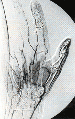
Preoperative brachial angiography was performed in 10 patients at our hospital, whereas the remaining patients were studied in other institutions and referred to us for operation. Usually, the most common site of flow blockage was at the level of the proximal interphalangeal joint, but lesions sometimes originated more proximally in the metacarpal arteries (Fig 1). Doppler ultrasound in most instances confirmed the physical findings. Blood gas analysis and spirometry were also performed, and FEV1 was less than 40°/O of predicted value in 3 patients. Associate conditions were hypertension in 10 patients, cardiac ischemia with history of angina or myocardial infarction in 3, mild diabetes in 2, and chronic obstructive pulmonary disease in 4. Thoracoscopic sympathectomy was performed under general anesthesia with double lumen intubation, with the patient placed in the thoracotomy position with the upper limb abducted and raised. Two or three entry sites in the axillary area were used: one in the V intercostal space for a 5- or-10 mm 0-degree scope. Instruments were inserted through the third or fourth intercostal space. Anterior rotation of the operative table was helpful in lung retraction. In 5 patients, diffuse pleural adhesion was encountered, and it was necessary to partially mobilize the lung to expose the sympathetic chain; in 4 patients, moderate thickening and opaqueness of the parietal pleura caused difficulties in identifying the nerve. To dissect the sympathetic chain, the parietal pleura was opened and the nerve was progressively isolated using scissor or hook dissector. The main trunk was prepared between T2 and T4, taking care to avoid lesion to the Stellate ganglion, and gently lifted up. This maneuver allows identification and resection of all collateral branches. Finally, the trunk was divided proximally immediately after the Stellate ganglion, distally at the level of T4, and removed. The nerve of Kuntz was isolated and resected, when evident. At the end of the operation, after accurate control of the hemostasis, a chest drainage was left in place and the skin incision was closed in the usual fashion. |
 |
|
Fig 1. Arteriogram demonstrates segmental obstructions in digital arteries and further lesions in palmar arch. |
||
Results
We
performed 10 unilateral and five bilateral staged (mean interval
time was 3 months) thoracoscopic sympathectomies. There were no
perioperative deaths. Two minor complications occurred: 1 patient
developed a pleural effusion after chest tube removal, which required
one thoracentesis; and 1 patient had an atrial fibrillation successfully
treated by anti-arrhythmic drugs. No Horner’s syndrome or
other collateral effects were noticed. Mean duration of postoperative
chest drainage was 2.5 + 0.4 days and mean postoperative hospital
stay was 5.3 + 0.5 days. Follow-up ranged between 3 and 71 months,
with a mean of 33 months. All patients demonstrated evident clinical
benefit after the operation: 5 patients had complete healing of
the ulcers and trophic lesions; and 6 patients had improvement
of their lesions and clear demarcation of necrotic areas. Doppler
ultrasound proved that blood velocity and local flow increased.
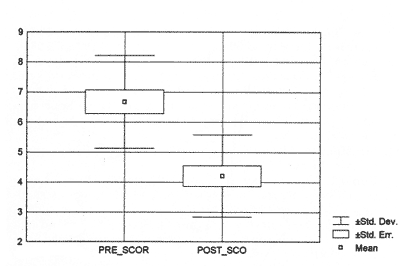
Reduction of pain was observed in all patients. Post-operative
severity of pain score was 4.20 + 1.54, and the improvement from
preoperative values was statistically significant (p < 0.05)
(Fig 2). Pain medication was suspended in 6 (40%) patients, reduced
in 6 (40%), and unchanged in 3 (20%) (Table II). Two
patients died during follow-up: 1 of myocardial infarction and
1 of cerebral ischemia, 24 and 32 months, respectively, after the
operation. |
||
Five
patients required one or more digital debridments and medications.
Four patients with tissue necrosis ultimately required delayed
amputation at one or more distal interphalangeal joints; in each
case, amputation was delayed until clear tissue demarcation developed,
which allowed maximal preservation of viable tissue. In the group
of patients with acute ischemia due to intra-arterial injection
of drug, 2 had a normal extremity. The remaining patient, who
developed tissue necrosis, required several digital debridments
and delayed amputation of a single distal phalanx; he also had
a permanent neurologic dysfunction with inability to flex the
distal phalanx of his thumb and weakness of grip.
|
 |
|
Fig 2. Comparison between preoperative and postoperative severity pain score (p<0.05). |
||
| BACK | TOP | NEXT |
|
 |
|